
Hisham Elseweifi
2023 Students
Biomedical Engineering
Department of Neurosurgery
Pronouns: He/Him/His
Email:
Week 1: Simple Does Not Always Mean Better Heading link
Good Design: Intraoperative Neuromonitoring system
Although there are less invasive variations, lumbar interbody fusions are nonetheless invasive procedures. During the surgery, there is still a risk that the surgeon’s hands and instruments directly contact the patient’s nerves, potentially resulting in permanent damage. Hence, a surgeon would benefit from a means to gauge that risk in real-time and how that risk changes with respect to each movement and interaction with a given patient’s spine.
Behold the Neuromaster MEE-2000 (Nihon Kohden) and the team that operates it.
Activities
The Neuromaster MEE-2000 has real-time SSEP, ABR, EMG, EEG, and TcMEP acquisition capabilities during the surgical procedure, otherwise known as intraoperative monitoring. These metrics provide a means to ensure the patient’s sensory and motor pathways remain intact.
Environment
The device was used in each of the three surgeries I observed this week, two transforaminal lumbar interbody fusions (TLIFs) and one anterior lumbar interbody fusion (ALIF). Its placement relative to the entrance and the patient’s orientation was nearly identical in the three operating rooms. The device is fixed in the corner of the operating room, facing the patient’s head, and directly next to the anesthesia team. This placement allows the technicians operating it to be well out of the way of any traffic. It gives the device’s electrical stimulators a safe pathway to the patient without presenting a potential tripping hazard. Moreover, as one of the technicians emphasized, specific actions taken by the anesthesia team may directly influence the Neuromaster’s readings; thus, the proximity of the two groups allows for immediate and seamless communication between them.
Interactions
The device was hooked up to the patient in a way to be able to stimulate and measure both the somatosensory and motor pathways. The physical configuration of the device’s components and the software used to operate the device allows for simultaneous visibility of most of the signals to the technicians. Additionally, the software enables the user to collect TcMEPs with the click of a button. The technicians also interact with the surgeons, alerting them if the readings indicate that the surgeons are dangerously close to the nerves or if the technicians want to collect TcMEP data. However, I also observed the surgeons requesting the technicians to collect TcMEP data before moving on to the procedure’s next step. Lastly, as mentioned before, the Neuromaster’s technicians and the anesthesia team interact to ensure accurate readings and avoid patient complications.
Objects
The Neuromaster MEE-2000 is a group of components working in unison. These devices include, but may not be limited to, a channel amplifier, acquisition breakout boxes, a TcMEP matrix stimulator, electrical breakout stimulators, monitors, and a keyboard and mouse.
Users
The technicians are referred to as the neuromonitoring team, or more formally, the intraoperative monitoring (IOM) team, and were the only ones I observed to operate the device directly. However, it goes without saying that surgeons are highly dependent on this technology and the IOM team to constantly monitor the patient’s sensory and motor pathways to help prevent devastating patient complications.
Bad Design: Surgical Screwdriver
Our task was to maintain an engineering perspective as we observed interactions within the operating room, NOT to walk out of there with an understanding of the surgical procedure. However, acquiring a high-level understanding of the general procedure allowed me to identify a design flaw and comprehend its potentially disastrous implications.
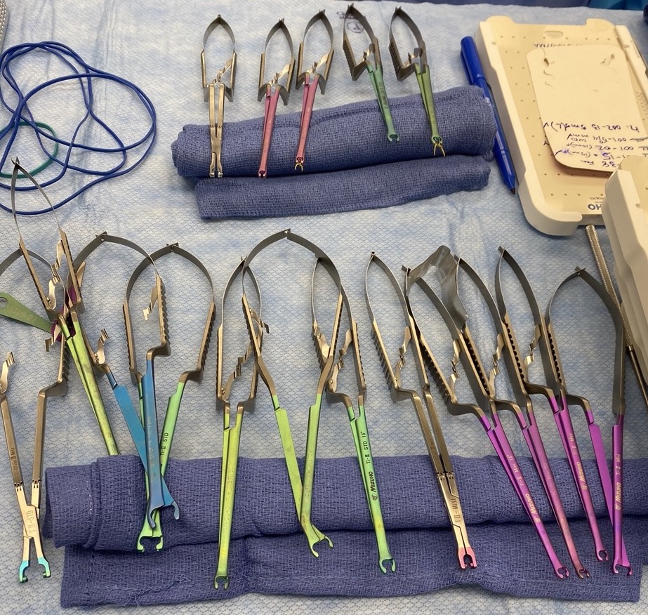
The surgeons were equipped with an assortment of sophisticated tools used to interact with and navigate the patient’s spine, as well as insert surgical implants such as screws, rods, and interbody cages.
The intriguing part was that a completely different vendor supplied their own assortment for each surgery. Hence, these tools were mechanically and ergonomically different from those used in the first TLIF, requiring the presence of vendor representatives in the OR during each surgery to instruct the surgeons on which tools to use and how to use them. Nonetheless, each toolkit was intuitive for the surgeons, and there seemed to be no real challenges—except during the second TLIF. While the surgeon had inconveniences with multiple aspects of the toolkit (notably the tower), the problematic design of the surgical screwdriver was the clearest. It will be of focus in this section. The screwdriver sacrificed being ergonomically friendly for functional simplicity.
Activities
At some point in each lumbar interbody fusion observed, the surgeon implanted pedicle screws for further spinal stability. Each vendor supplied specially-designed surgical screwdrivers used to insert these screws safely. The surgeon would twist with his right hand and guide the driver with his left hand.
Environment
I am only aware of this specific screwdriver being used for TLIFs. The screwdriver came sterilized in a package and was unsealed and positioned on the tool table once the surgery began. The table was within reach of the medical student assisting the surgeon, ready to be handed to them when required. Moreover, the table was entirely within the view of the vendor representative. It is also important to note that an exemplary aspect of the driver’s design was its compactness; hence it occupied little space within the incision.
Interactions
When it came time to use the driver, the flaws associated with its simple design became apparent. As stated above, the surgeon preferred placing his left hand in the middle section of the driver to guide the tool while his right hand operated the ratchet mechanism at the top of the driver. However, he quickly discovered that he was, in fact, disassembling the screwdriver while attempting to drive one of the pedicle screws into the bone. The issue was that the locking mechanism was also placed in the middle of the screwdriver, and the same motion used to drive the screw in the bone was used to assemble (and disassemble) the handle to the driver shaft. According to the vendor representative, instead of wrapping his hand around the middle of the shaft as he was accustomed to, the surgeon was supposed to use his left hand to grip the bottom of the shaft using his index finger and thumb, similar to holding a pencil. The surgeon did not find this ergonomically friendly but could still successfully carry out the procedure without complications to the patient.
Objects
The screwdriver had a two-part design consisting of the driver shaft and the handle. Different handles were available to suit the user’s preferences and needs. This design was allegedly simple and intuitive, but the forced repositioning of the guiding hand did not make this a favorite for the surgeon.
Users
Although the neurosurgery residents performed select steps of the surgical procedure, I only recall seeing the head surgeon utilize the screwdriver. However, as noted above, the driver was handed to the surgeon by the assisting medical student, and the vendor representative provided instructions.
Week 2: Introduction to Storyboarding Heading link
Week 2 of clinical immersion introduced us to new domains of the neurosurgery department. We started the week in the Neuro ICU with professor and chief of the neurocritical care division, Dr. Ramos-Estebanez, who brilliantly complemented each patient examination with an interactive discussion to ensure an educational experience for everyone—the following two days entailed new surgeries, most notably an endoscopic TLIF, a corpectomy followed by a posterior cervical disc fusion, and a craniotomy with a vertebral artery aneurysm clipping. Lastly, Friday again allowed us to appreciate the clinical aspect of surgical care as we shadowed Dr. Mehta’s pre- and post-operative interactions with patients.
This week we were tasked to home in on one observed procedure, provide a step-by-step illustration, and identify points that potentially complicate each step. Of each process observed this week, surgical and non-surgical, I found the craniotomy with aneurysm clipping to be the leading candidate due to the technologies that drove its execution. The clipping portion of the surgery was an entire mission alone. Hence, I limit the discussion to the attending neurosurgeon’s perspective of that portion of the surgery, from when the aneurysm was located to when it was clipped correctly.
1. Temporary Clipping of Aneurysm
The aneurysm was located on the patient’s vertebral artery and was within view of the microscope by this point of the surgery, but its accessibility was questionable. Moreover, before the surgeon attempted to clip the aneurysm, he placed a temporary clip a bit further down the artery from the aneurysm. According to Kumar et al., temporary clips are an often crucial, preemptive step in aneurysm clippings, as they act as a prophylactic measure to intraoperative aneurysmal ruptures during aneurysm clippings. However, the authors also noted a consequential increased risk of ischemia due to prolonged use. The nerves and blood vessels seemed to be obstacles to the temporary clip, as the artery was deep within the exposure. These obtrusive structures appeared to be at risk of being struck by the clip applier throughout the entire aneurysm clipping procedure.
2. Determining Approach
The aneurysm was manipulated as necessary, but its position, orientation, and geometry remained an issue. The orientation was an issue because the clip applier tool could only be angled as a whole and from the surgeon’s wrist; no point along the tool’s length could be adjusted, bent, or rotated. Given the aneurysm was not facing up perpendicular to the entry point but was instead angled, the surgeon had to tilt his whole wrist and hence the entire clip applier to place the clip. As a result, the surgeon had access to many clip appliers and clips of various shapes and lengths. This meant the risk of bumping into the previously mentioned obtrusive structures and the brain and spinal cord.
3. Applying permanent clip
The surgeon found a safe path to the aneurysm with the clip applier, but now it was a question of which clip to use. The geometry of the aneurysm neck was wide and hill-like. Every single clip the surgeon tried to apply ended up sliding off. The surgeon attempted at least three different clips, each facing this problem. When he finally got a clip to stay, the ends of the clip blades did not come into contact. Displeased with the incomplete clip closure, the surgeon attempted to apply a second clip facing the opposite direction but did not have luck.
4. Verify Successful Occlusion
The operating team decided it was time to verify if the aneurysm was successfully clipped. The microscope was equipped with a monitor, which also transmitted its display to the other screens in the room throughout the procedure. Everyone could see what the surgeon could see. The microscope also featured a near-infrared indocyanine green video angiography (NIR ICG VA). The fluorescent dye was inserted, and the display revealed it flowing through the artery, but none entered the aneurysm. After prudent review, they concluded that the aneurysm was clipped. Nonetheless, the surgeon had to wait at least 20 minutes after placing the clip to test the patient’s motor function before closing.
One issue I observed regarding the microscope was it was unbalanced and uncalibrated for a good portion of the surgery. The surgeon had a different field of view from the fellows and residents observing through the monitor, and the microscope (which was stabilized with the surgeon’s mouth) kept moving during the surgery. This frustrated the surgeon, who had to request a tech to recalibrate and reposition the device.
5. Disengage and Close Exposure
After replaying a recording of the ICG VA to verify the aneurysm was successfully occluded and testing to ensure the patient’s motor system was intact, the surgeon was ready to disengage. First, he had to remove the temporary clip and ensure no other damage was done. Afterward, it was closing time.
Final Thought
Interested in the success of ICG VA, I browsed the literature for some reports of the intraoperative imaging technique used in other aneurysm clippings. According to a review from Balamurugan et al., although many studies have demonstrated consistent success in using ICG VA to verify aneurysm occlusion, multiple studies have found misleading results from ICG VA. The authors concluded that even if an ICG VA shows complete occlusion, residual flow can still occur following the procedure due to incomplete closure of clip blades and aneurysmal atheroma. On the other hand, another study found that repeated injections of the dye too frequently to one another could result in type 1 errors.
References
Balamurugan, S., Agrawal, A., Kato, Y., & Sano, H. (2011). Intra operative indocyanine green video-angiography in cerebrovascular surgery: An overview with review of literature. Asian Journal of Neurosurgery, 6(02), 88–93. https://doi.org/10.4103/1793-5482.92168
Kumar, S., Sahana, D., & Menon, G. (2021). Optimal use of temporary clip application during aneurysm surgery – In search of the holy grail. Asian Journal of Neurosurgery, 16(02), 237–242. https://doi.org/10.4103/ajns.ajns_465_20
Week 3: Needs Statements — Who Needs What? Heading link
This week focused on the development of needs statements. At the heart of user-centered design, needs statements offer a structured, coherent, yet iterative framework for identifying problems without implicit bias toward a solution. Considering the formal introduction of this concept was a first for many of us, our exercise this week was to develop three iterations of one needs statement while elaborating on the population, opportunity, and outcome.
Our team identified several themes of interest, but this blog post will focus on a particular recurring issue in just about every spinal surgery we have observed: surgeon familiarity with surgical equipment. From day one in the operating room, I was in awe of the surgeon’s ability to adapt to surgical equipment, which he had never been exposed to at the instructions of vendor representatives in the OR. Aside from the surgeon’s unsurprising intuition in operating the tools, the rigorous training and education received by each vendor representative in the operation and application of each instrument is vital for this process.
I was recently informed that neurosurgeons are generally invited to conferences to try these tools a few weeks before using them in operation. Despite this, the assistants who retrieve and assemble the instruments during the surgery are not typically invited and, thus, often need to be more familiar with the equipment. The vendor representative must gauge the required tool, point it out to the assistant, instruct them on assembling it, and then explain how to operate it to the surgeon. Additionally, surgeons can’t be expected to retain each detail in working the equipment multiple weeks after a conference. Moreover, the vendor representatives are not medical doctors, let alone trained neurosurgeons. Problems may arise if the surgeon must diverge from the traditional procedure. I had witnessed instances where the vendor representatives were unsure what the surgeon was trying to do when the surgeon himself was unsure how to use the tools he had to accomplish a particular step of the procedure.
With all that said, who needs what?
1. Neurosurgeons operating new surgical toolsets require new approaches to gaining familiarity to reduce operating time.
Population: Neurosurgeons operating new surgical toolsets
Opportunity: Gaining familiarity with said toolset
Outcome: Reduce operating time
As an engineer, I would be lying if I said I don’t already have three different solutions to this problem I am aching to explore. Refraining from any implication of a solution in this needs statement is rather challenging, and I am admittedly struggling with the opportunity aspect of the message. However, in placing all my focus on that, my bias still found its way to the population. The winning solution may target the assistants or the representatives, or it may encompass all three. Moreover, perhaps near-identical scenarios occur in surgical operations outside of neurosurgery.
2. Reduce the knowledge and language barriers of surgical tool operation between surgical equipment representatives, operating surgeons, and surgeon assistants to reduce operating time and minimize the risk of surgical complications.
Population: Surgical equipment representatives, operating surgeons, and surgeon assistants
Opportunity: Knowledge and language barrier of surgical tool operation
Outcome: Reduce operating time and minimize the risk of surgical complications
The opportunity seems now too unclear. Also, I can phrase this in a way that still encompasses all three parties but offers more direction.
3. Vendor representatives require supplements to aid in familiarizing surgeons and assistants with their equipment during surgeries to reduce operating time and minimize the risk of surgical complications.
Population: Vendor representatives
Opportunity: Operating team familiarity with new surgical equipment during surgeries
Outcome: Reduce operating time and minimize the risk of surgical complications
From my interpretation, this supplement can be a process or a device, and the familiarizing can occur before or during the surgery. I could have said, “Surgeons require supplements to aid in familiarizing themselves with the equipment…”, but the presence of vendor representatives in the OR is a means to solve the unfamiliarity problem. There is no downplaying the importance of the vendor representative’s role in the operating room, and I don’t imagine them being replaced anytime soon. Likely, the vendor representative will have a role in implementing solutions to the problem, even if they don’t directly use it to familiarize the surgeons and assistants.
Still, there is room for improvement, and maybe in a week, I will end up swapping the population back to the surgeons. I will also investigate how relevant this issue is in other surgical departments. Regardless, I certainly greatly appreciate needs statements after this exercise.
Week 4: Intellectual Property Heading link
Following a review of the implications of intellectual property law in device design and manufacturing, Dr. Browne and Dr. Felder tasked us with considering the role of patents in our examination of medical devices this week.
With our mentor out of town, I had the opportunity to observe another aneurysm clipping case.
This time, the aneurysm was located along the right posterior communicating artery with vastly different geometry and orientation. The geometry didn’t seem to pose as much of a challenge during this case, as none of the clips slid off. On the other hand, the unclear position and orientation of the aneurysm led the surgeons and residents to bounce back and forth from the angiograms to the microscope display to identify the surrounding structures and verify the surgeon’s location. Once the team felt confident they were in the right place, the surgeon attempted several clips before settling for a fenestrated angled clip to wrap around the parent artery without requiring further exposure or maneuvering.
Afterward, the operating team employed ICG to verify successful occlusion. However, given the position and orientation of the aneurysm, it didn’t seem possible to thoroughly evaluate the situation based on the ICG VA alone. As touched upon in a previous blog post, the ICG VA display was integrated with the microscope as part of one whole surgical visualization device (Zeiss Kinevo 900). The challenges presented by the orientation and position of the aneurysm also made me think back to Medtronic’s Stealth station computer-assisted navigation system I observed being used for a minimally invasive T12-L2 fusion last week. I was curious if any designs have been conceptualized to encompass navigation and visualization into one device to optimize and enhance surgical procedures similar to the one I observed that day.
Indeed, a very recent patent was published:
“WO2023039596 – Integrated surgical navigation and visualization system, and methods thereof”
In fact, this patent mentions both models as it clarifies the distinction between surgical visualization and navigation. The patent seems to make claim to a device that encompasses many of the optimal features of the Zeiss Kinevo 900 visualization system, namely “single cart providing mobility”, “an N-camera stereoscopic digital surgical microscope, wherein N is 2 or greater”, “an orientation adjustment handle”, and several others. However, the key concept here is the unification of a navigation system with a visualization system as one single device. According to the patent, discrete visualization systems already exist that can integrate with other navigation systems, but such is still a system of two different units. This patent claims a system with a single unified user-responsive display that provides both navigation and visualization of the surgical site. The description seems to elaborate that there may be more than one display on the device, but at least one display incorporates the above-mentioned integrative feature.
I am curious how this patent has been acted upon and how such a device could have reimagined this week’s aneurysm clipping case.
https://patentscope.wipo.int/search/en/detail.jsf?docId=WO2023039596&_cid=P22-LKQD9D-86630-1
Week 5: Market Assessment Heading link
With CIP concluding, my team and I had to narrow down our selection of needs statements to pursue. We spent one last week observing cases in the OR and brought our final set of needs statements to our mentor, Dr. Mehta, a neurosurgeon. Though our discussion left us still with a few needs statements, ultimately, they all revolved around problems neurosurgeons encounter when carrying out endoscopic spinal surgeries. Dr. Mehta also informed us that an incredibly small fraction of neurosurgeons perform endoscopic spinal surgeries; the number may be as low as 15 neurosurgeons. However, Dr. Mehta anticipates this number to grow exponentially in the coming years.
One of the problems regards being able to identify in real-time the exact location of the endoscope relative to other structures in the body. A needs statement for that would go something like this:
Endoscopic spinal surgeries require a method of real-time visualization of the endoscope’s position relative to patient anatomy to minimize unnecessary invasiveness, reduce operating time, and influence more neurosurgeons to adopt minimally invasive methods.
As this statement stands, the target population solely encompasses neurosurgeons. This scope may need to be narrower as a potential solution may integrate seamlessly into endoscopic surgical practice outside neurosurgery. However, further research is required in order to verify this.
With that said, I wanted to perform a rough market analysis pertaining to a pursuit of this needs statement, namely an estimate of the total addressable market (TAM), which is computed as:
(Number of units per year) * (Cost of product)
Unfortunately, the total number of each type of endoscopic spinal surgery performed each year, as well as their associated costs, were not readily available. According to the Agency for Healthcare Quality and Research National Inpatient Sample Database, at least 735 endoscopic spinal surgeries were performed in 2020, though this number only accounts for discectomies and spinal fusions. UIC MyChart lists out-of-pocket spine fusions as an estimated $32,272. From these numbers alone, the TAM would be calculated as ~ $23.7 million per year. Of course, however, this is erroneous even as a rough estimate. This estimate does not account for the potential cost variation with each type of endoscopic spinal surgery (i.e., spine fusion vs discectomy vs laminectomy).
Moreover, the cost almost certainly factors in (or maybe only accounts for) open, as opposed to minimally invasive, surgeries. Lastly, not only is there an anticipated rapid growth in the number of neurosurgeons who can perform these surgeries, the scope may include other surgeons. Hence, $23.7 million per year is not a meaningful estimate. Further identification of specific procedural costs could help to at least establish a minimum TAM, as the limited available information hinders a truly accurate estimation.
Week 6: Concluding Remarks Heading link
CIP was a career-defining experience, to say the least.
Not including Monday’s educational workshops delivered by Dr. Browne and Dr. Felder, we spent over 120 hours shadowing our mentors and other physicians throughout the program. My team and I observed more than 15 neurosurgeries in the OR, both spinal and open brain, performed by Dr. Mehta, Dr. Atwal, and the neurosurgery residents. Additionally, we spent over 30 hours shadowing our mentor, Dr. Mehta, through his pre- and post-operative interactions with patients in the clinic. Finally, we followed a couple of different neurologists and neurology residents as they carried out rounds in the Neuro ICU.
To all future biomedical engineering participants:
- While there is no question that the medical underpinnings are intriguing, observe the procedures through the lens of how you can innovate. Otherwise, do not get too distracted or worried about understanding every detail of the process through observation. There is too much to digest, which the med students will be more likely to comprehend. You can also ask relevant questions to your mentors or do some digging online.
- Talk to the vendor representatives (red and dark blue scrubs) and other operating team members as early as the first surgery! Many are sufficiently knowledgeable about the procedures and associated technologies and will happily share their knowledge. Moreover, befriend the other present med students outside of the CIP program.
- Take notes! Documenting your observations and critical technologies used is unbelievably valuable. This includes the manufacturer name, device model, associated procedure, and other details. Ask the vendors how the devices work and if they can provide any more supplemental information or sources. Also, take photos as permitted!